Restorative dentistry
Considerations regarding the actual extent of a grade 3 Interproximal Carious Lesion
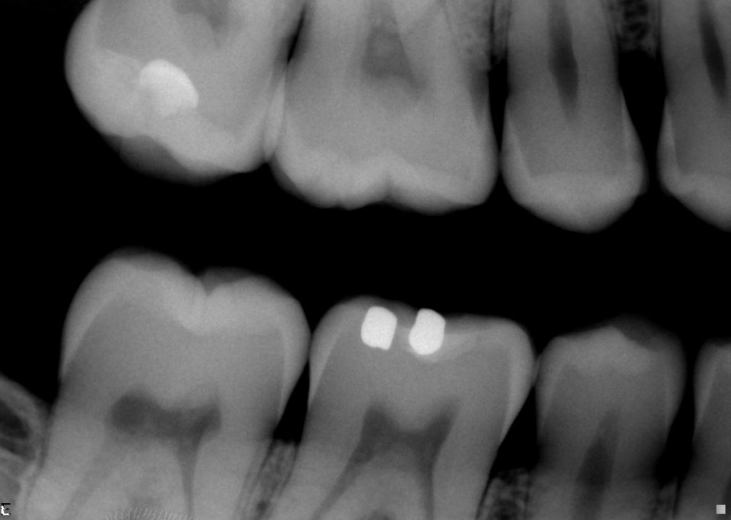
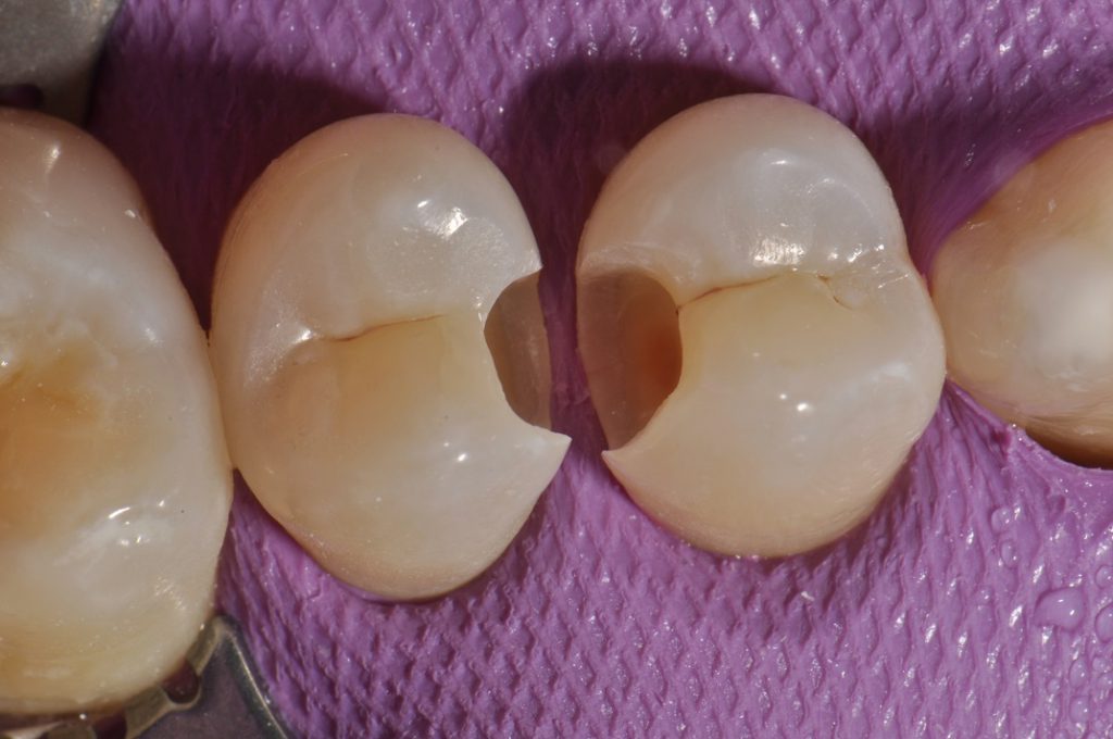
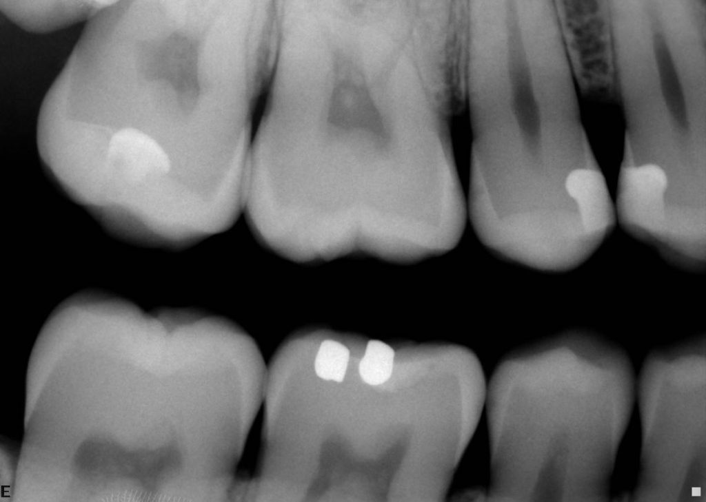
I have heard from a certain source that some students are being taught that Grade R3 carious lesions (cavities that just peak past the dentinoenamel junction (DEJ) radiographically into dentine) can be in the right conditions self-limiting and reversible. I guess this is possible if you can change the oral condition from that of being ‘demineralising’ to one of remineralisation and/or if you can seal the lesion or decalcified enamel channel feeding it. This case demonstrates the unexpected extent of such a lesion especially on 15MO. This is a lesion which might be “watched” ‘by a clinician but is actually quite deep in the dentin clinically. Also, there is no real way to “seal” off an interproximal lesion predictably, although there are restorative technologies such as resin infiltration by DMG Icon that appear to show promise.
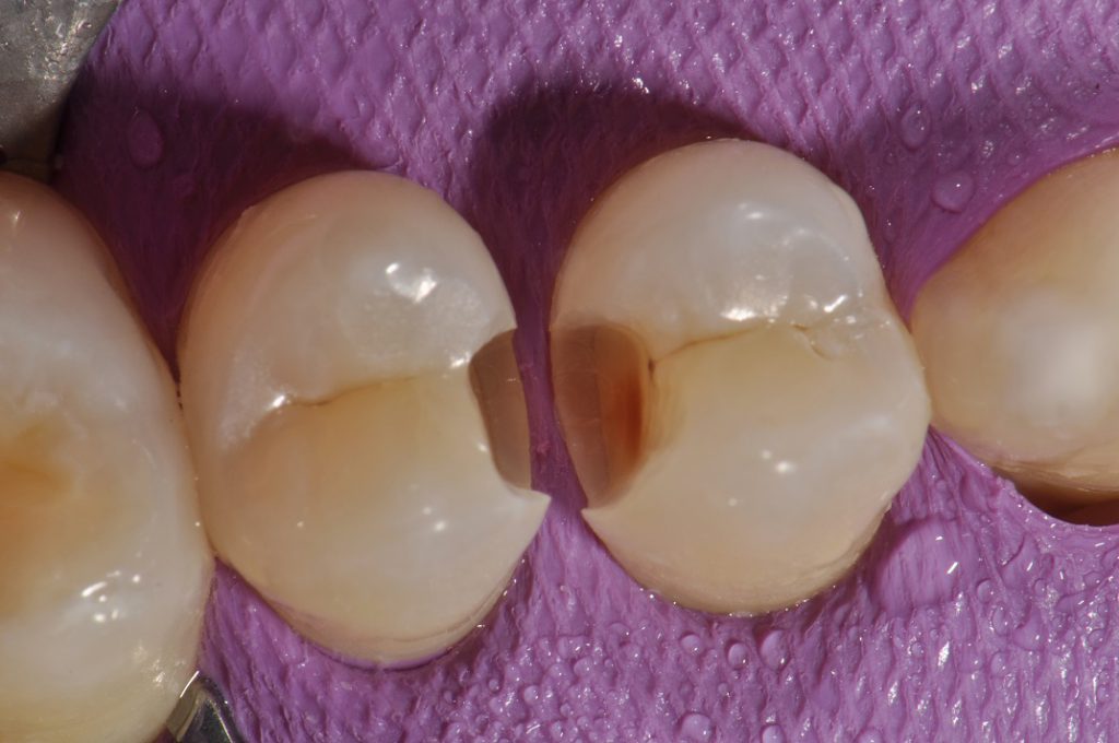
In addition to the 15 mesial R3 carious lesion, the 14 distal R3 lesion was definitely extended into the outer half of the dentine. After initial preparation, you will see the further extent of demineralized and discoloured dentine, which was also soft, extending pulpally.
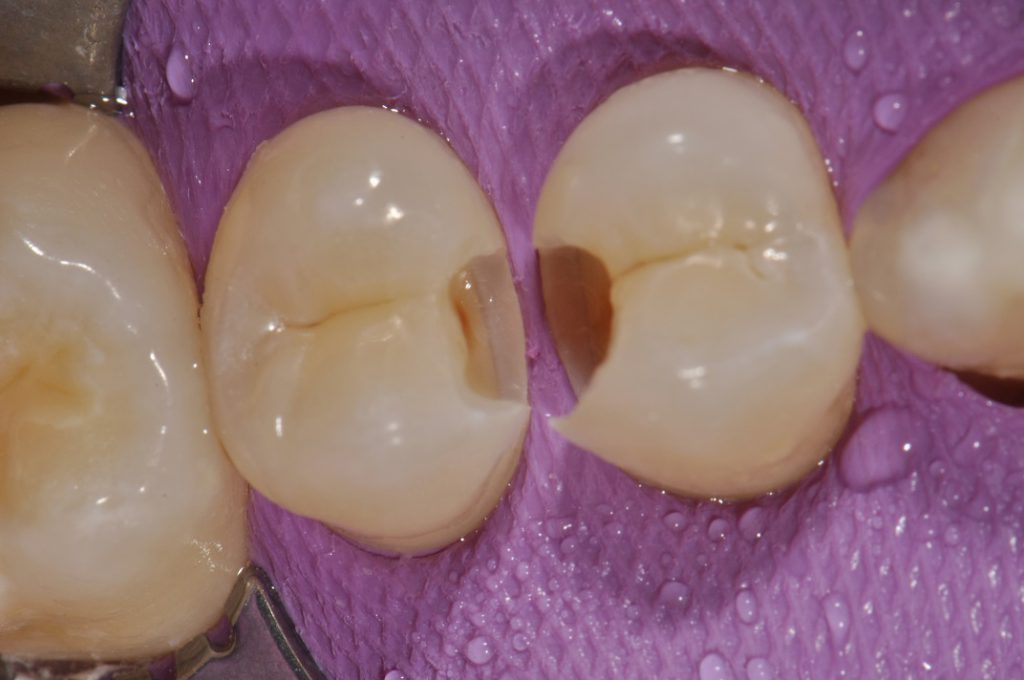
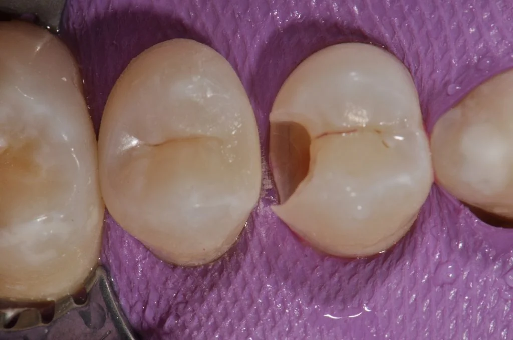
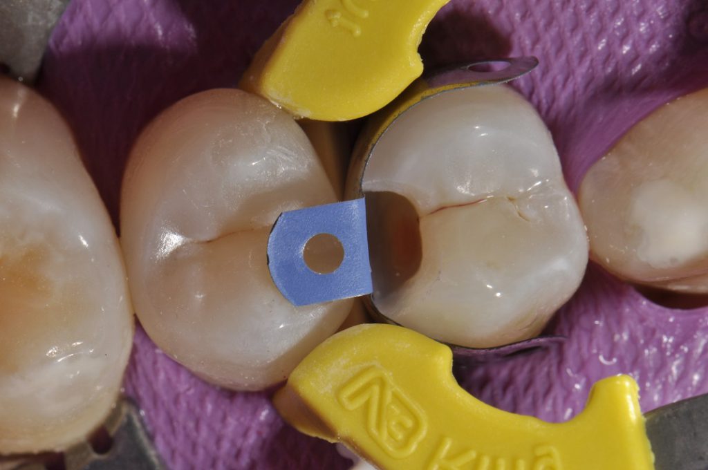
Stained or discoloured dentine is not always infected dentine. On excavation with the slow speed round, you can see the next two photographs exhibit the lesion in mid-preparation and in final outline form after removal of all affected dentine. This was assisted via use of a caries detector dye (Kuraray) and slow speed round bur excavation revealing the “true extent” of the caries. The proximal wall of the 15MO was restored with the help of an Omnimatrix and shaping this wall before applying the Dentsply Triodent V3 sectional system to the 14DO (a 5.5mm SuperCurve non-stick matrix was placed before the medium Pink Wave Wedge). Following this, a yellow, premolar-sized V3 tensioner ring was secured interproximally with the tines straddling the Wave Wedge. The contact area was burnished with a ball burnisher. Following
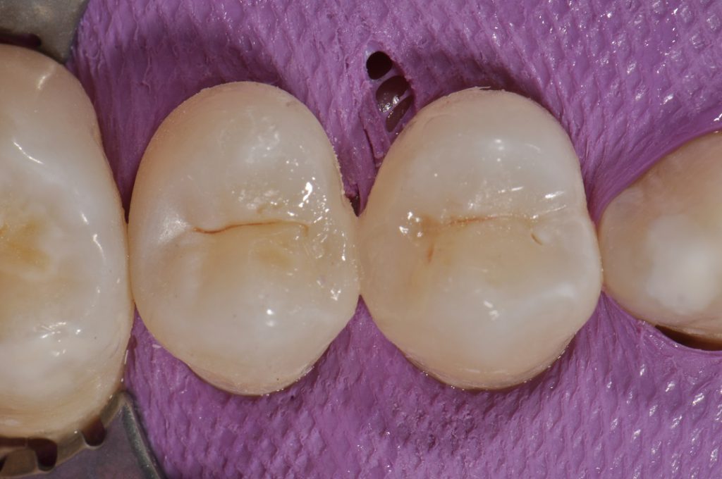
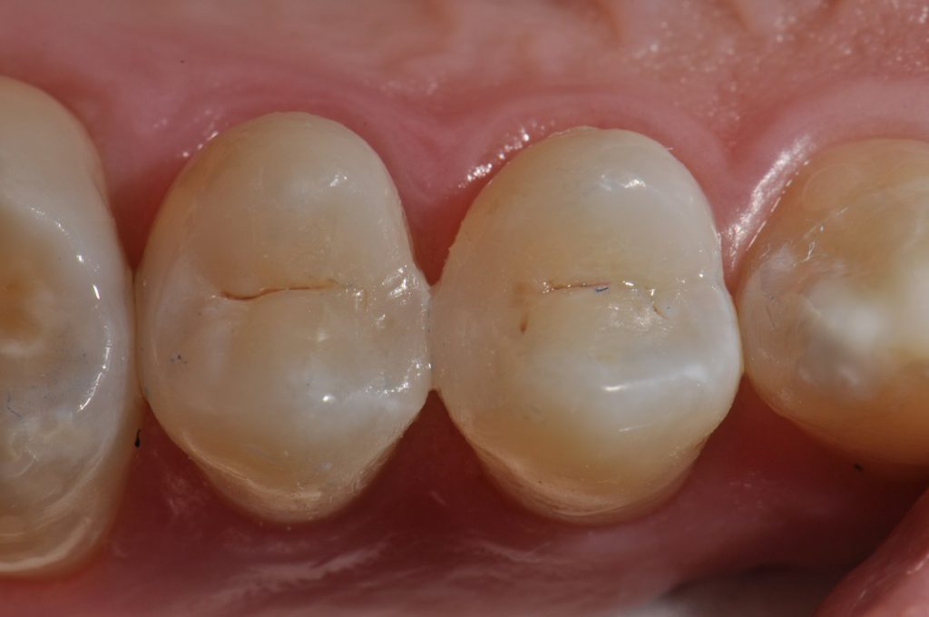
An effort was made to recreate the slight mesiolingual concavity on the proximal wall of 15, a little light stain (GC Optiglaze Colour Kit: Pink Orange) was touched into the fissure re-creation.
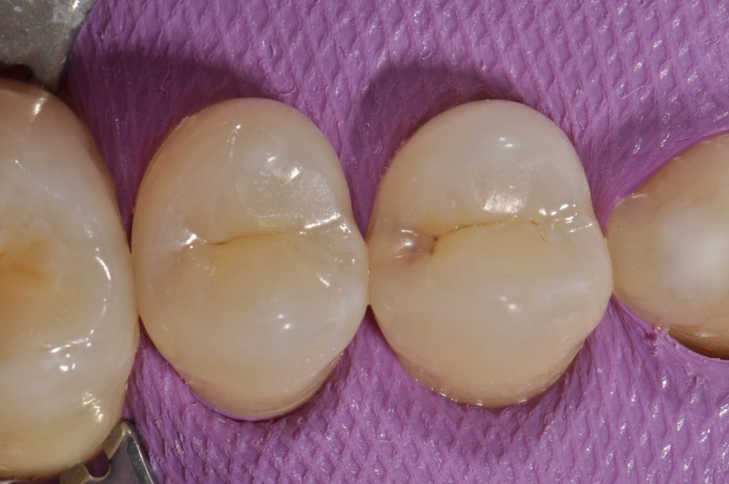
The entire restoration was completed with a single shade of GC G-Aenial Posterior A2 following a thin liner of GC G-Aenial Flo X A2, to achieve an outstanding chameleon effect.
The post-operative film is attached for comparison. Early Grade 3 lesions “are” dangerous. The radiographic extent of R3 carious lesions are not always predictably confined to the outer dentin, as a lesion is only visible radiographically when 1/3rd of the buccolingual width of the tooth is affected. (BDJ 1988; 164(80):243-247).
Often, occlusal and some proximal carious lesions tract narrow and deep, as they do in some deeply stained fissures and marginal ridge hairline fractures with no radiographic evidence of dentinal involvement. My personal clinical approach is to treat R3 lesions to prevent possible more extensive tooth destruction in the future. Food for thought. If you have access to DMG Icon, you can definitely try to remineralize the lesion, although the final result is not radiopaque. You will need to follow the patient and the radiographic lesion closely on recall examinations.